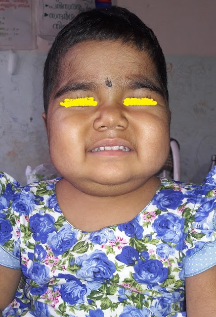
Four year old girl child presented with fever cough and dyspnea. No running nose ,cough was dry .
She was hospitalized and was on Inj Ceftriaxone.
As the dyspnea worsened she was referred to us.
She was hospitalized and was on Inj Ceftriaxone.
As the dyspnea worsened she was referred to us.
First child born at term out of uneventful antenatal history was not asphyxiated. She had neonatal convulsions and was hospitalised for twenty days ? meningitis.
Development was normal .
Last year she developed thrombocytopenia ,diagnosed and managed as ITP .
There is history of abscess drained three times from different sites. Immunized update.
No family history of any significant illness. ( No consanguinity) . No contact with tuberculosis.
Development was normal .
Last year she developed thrombocytopenia ,diagnosed and managed as ITP .
There is history of abscess drained three times from different sites. Immunized update.
No family history of any significant illness. ( No consanguinity) . No contact with tuberculosis.
No past history of recurrent respiratory infection,ear discharge,purulent nasal discharge ,loose stools .
On admission she was dyspneic, fully-conscious, maintaining saturation above 90 percent with 50 percent oxygen circulatory status stable .
No focus of pyoderma or abscess.
No rash or bleeds,
No Lymph nodes
No stigmata which grossly suggest inherited immunodeficiency .
No focus of pyoderma or abscess.
No rash or bleeds,
No Lymph nodes
No stigmata which grossly suggest inherited immunodeficiency .
Her weight , height and Mid arm circumference was adequate for her age .
Respiratory system exam
Upper airway normal
Chest movement diminished on the right side.
Apex and trachea shifted to left. Percussion stony dull in the right lower inter scapular axillary infra mammary areas. Air entry diminished same areas VR decreased ,no bronchial breathing, no added sounds.
Apex and trachea shifted to left. Percussion stony dull in the right lower inter scapular axillary infra mammary areas. Air entry diminished same areas VR decreased ,no bronchial breathing, no added sounds.
CVS normal except shift of apex to left
P/A no organomegaly, no free fluid
CNS fully conscious, no neurological deficit no Signs of meningeal irritation
In short
Four year old child admitted with fever ,dry cough and progressively worsening dyspnea.
Normal upper airway and mediastinal shift brings pathology below tracheal bifurcation. Mediastinal shift to left and stony dullness argue collection inside right pleural cavity. In view of fever in this age group most common reason is bacterial pneumonia with syn-pneumonic effusion. But here the cough is dry only and more features of fluid in pleural cavity than parenchymal involvement. Possibility of empaema considered high .
Is there an immunodeficienty behind ?
Teaching points for PGs at this stage of history .
Multiple abscesses drained ,and now empaema possibility of susceptibility to staph infection high ? Yes we should investigate in that line. Immunodeficiency states with abscess formation are
1. Problems with phagocytosis of neutrophils
2. Problems with respiratory burst ,chronic granulomatous disease. Usually boys but girls may be affected
3. Jobs syndrome ,
other entities also
Neutrophil related problem and bacterial infection . It may be adhesion problems , chemotaxis , phagocytosis or respiratory burst .
In problems with adhesion , and chemotaxis, neutrophils wont reach the site of organism . So NO evidence of inflamation, no pus . Common reason for this state is any significant neurtopenia. ( No neutrophils to reach the site ) . In adhesion problems usually the neutrophil count in the peripheral blood ll be high ( They wont go out ,but remain inside )
Course
USG confirmed empaema right side and ICD done .She was put on inj CP , Cloxacillin 200 mg/kg /day and Gentamicin .
Temperature touched baseline in two days .She was active ,playful .Exam showed trachea and apex in normal space ,dullness in the right infrascapular area , decreased VR and breathsounds same area.
So some more of pus remaining ?
Repeat Xray chest and USG was done
On the right side costophrenic angle was clear ,patchy pneumonia middle lobe. No abscess .
On the left side there was a circular translucency .
Two questions remained
1.What is the reason for the Clinical findings in the right infrascapular area. Xray and repeat USG did nt show fluid .So this was attributed to pleural thickening (not a good explanation as this usually it takes two weeks )
2.What is the translucent shadow on the left side.?
Possibilities for this shadow
1. cavity inside parenchyma .
2. Pleural adhesions
3.abdominal viscera
4.Pericardial cyst
Out of this possibility of abdominal viscera was less likely. ( diaphragmatic hernia and eventeration are the common ones , but here diaphragm is seen in normal place . Other possibility of rapture is less likely as there is no trauma involved.
This location pericardial cyst unlikely ( this large cyst anterior to heart
So main DDs considered were a partially filled abscess inside parenchyma and pleural adhesions causing an appearance like this .
If it is parenchymal it must be in the lower lobe.
If pleural it can either be in the front or back
The line of diaphragm could be traced medially as there was a confusion whether this represented level of fluid. One more argument against abscess was the translucency of air ( above the fluid, if it is an abscess ) was not dark enough.
So
Repeat X ray AP and Lateral was taken
So Now ?
There is no shadow in the lower lobe area as we postulated. The shadow in the mid zone represent the middle lobe consolidation. About the shadow we were considering is in the anterior part , and the level is clear , traceable beyond the diaphragm . It is a partially filled abscess.
Rechecked for clinical findings in the same area. Normal resonance in percussion ,Normal intensity of vesicular breath sounds ,normal resonance .
She remained a febrile ,active except for the fact that pus was draining 40 to 50 ml per day . Air was bubbling episodically .
As there is possibility of bronchopleural fistula and lung expanded well we did nt ask her to inflate balloon which we used to do .
As she remained afebrile ,not sick not dyspnoeic same antibiotics were continued .We were curious from where this air and pus was coming as the X ray and USG did nt show any collection. As there was doubt whether there is chance of air entering to the tube from outside by accidental withdrawal , we checked for that also , and ensured ICD tube has entered enough .The air column was moving free .
Two other things which came in to consideration
1. Should we instill streptokinase ?. We used to and had good results. In this case we did nt . Because there is bronchopleural fistula and we were afraid whether this will affect the sealing of fistula .
2.Second point . Should we go for VATS. Yes . Many institutions resorts to early VATS and good results. Here in our institution it is being done, but not many cases and not for these young ones . We discussed with parents about the option to send to higher institution , but they opted to continue treatment here
On the tenth day of ICD the bubbling was significant .
There was argument for and against stepping up antibiotic . Consensus was to step up.
What were the arguments for continuing same drugs . Kid is not sick , not febrile . Lung findings were not worsening ,xray and USG shows clearance. The shadow in the left side also does not need any intervention apart from what she is getting .
Opposite view was..Patient is already on ten days of antibiotics. Pus still coming , broncho-pleural fistula is there. Is it safe to continue first level antibiotics ?. Yes staph is always dangerous. ( One point forgot to mention that culture did nt yield growth )
We decided to step up
Next question , stepping up to which one. Vancomycin ? Linezolid Clindamycin ?
Vancomycin is superior for MRSA ,but inferior to cloxacillin or other antistaph agents if not MRSA .In this case chance for MRSA is less. She responded very well to first line . Spectrum of action of vancomycin against other organisms less .
Next choice was between Linezolid and clindamycin. Our experience with Linezolid for the last few years were excellent ,Good response most of the kids tolerating the drug very well .
She was put on Linezolid 10 mg per kg per dose three times daily. Today fourth day of Linezolid. The air leak stopped completely, the pus drainage reduced. No pus yesterday. We were planning to remove the tube today . But today ten ml of pus still there. Now we decided to continue ICD for few more days .
What are our plans now
1.First control present problem
Next step to find out the reason behind , ie there is immunological basis . We are planning to do the immunoglobulin levels. We had hyper igE syndromes presenting like this. One of those cases now going well with long term co trimoxazol orally low dose .
If results are negative, may need to consider rarer entities.
Shall let you know the follow up
8/8/2016
On sunday she developed sudden dyspnea. Xray was taken and showed this
ICD was blocked ,resulting in pyopneumothorax .ICD changed and distress releived . Fifty ml of pus drained .
Linezolid stopped and she was put on Vancomycin and cefepime .
Next day draining of pus almost nil but the air bubbling continued, with each breath .
In view of this CT was done
;
Broncho pleural fistula .
ICD was continued for three weeks. Gradually bubbling stopped . We could remove ICD ,patient afebrile , about to shift her to ward
She developed sudden dyspnoea cyanosis
Xray taken
Again ICD was put
This time her dyspnea did nt improved. Bubbling continued.
We decided to surgical intervention . Patient opted for Surgery from Another institution
20 , december
Middle lobectomy done .
Specimen
Post surgery Xray
She is doing fine now


