14 year old girl, who had ASD closure five years back, was asymptomatic since then. Normal girl going to school, she was bit depressed as she had deformity of spine which was noticed in early childhood but later progressing. She now complaints of chest pain not related to exertion, mostly at night .Pain is vague she points to left side .She does not have dyspnea on routine activities. As she is having the deformity she does not participate in competitive play activities. No palpitation.
O/E
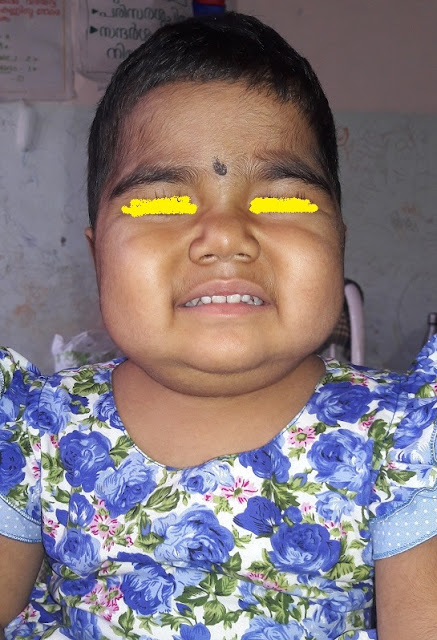
She is short 146 cms, no shortening of neck or webbing. Apart from deformity of spine, and shoulders no features suggestive of any syndrome.
CVS
Pulse 40 per minute, regular .high volume .
Blood pressure 100/70 in upper limb.
Did not record in the other limbs .
All peripheral pulses palpable.
(At this stage , two important points in the history …1.She is not on drugs .2. Bowel habits ,recent school performance ,3 Head ache vomiting .No need to explain the relevance of these questions in this context )
JVP not raised in her case
(Again , JVP analysis is important in this context .This low pulse rate , after ruling out other reasons we consider cardiac origin . One of the reasons for this low rate is complete heart block. JVP will show giant “a “waves episodically .Giant a waves seen with all atrial contractios when there is a mechanical obstruction to the RA eg Tricuspid atresia .
This video in you tube is another case , not this girls . Just to stress the usefulness of this examination in bradycardia
Precordial exam. Median sternotomy scar .Apex normal ,Heart sounds normal intensity ,Second sound wide split and fixed.Here again one interesting point. Second sound after surgical correction split remains as such . We all learned about the reasons for wide fixed split in ASD .Major reason we learned is the RA to RV contribution of volume is more than normal leading to wide split , and same volume irrespective of the phase of respiration . If that was the only reason , it should have disappeared once we close the ASD .But that does nt happen . So wide ,fixed split of second heart sound is not due to this alone.
There was an ejection systolic murmur ,grade II .at the pulmonary area . There was no third heart sound ,no clicks.
Why i said about all the above. You expect the systolic murmur also to disappear once the extra amount of RV outflow ceases with closure of ASD . That again does not happen. Why i said about third heart sound.? RV Third heart sound in an ASD indicates and helps to quantify the shunt , You dont expect that in a corrected situation , If it is there there must be another reason .The other reason for mentioning , We may get a residual lesion after closure. or an associated defect like small VSD or PS.The importance of mentioning Ejection click are two 1. Associated PS is so common in ASD ,If it is valvular there can be thrill , higher grades of murmur at second space and a click .One of the commonest association of Secundum ASD is mitral valve prolapse
Her other systems were within normal limits .
.

Chest Xray PA view . (some artifacts )

v5 v6

AVF &V1
Narrow QRS complexes. <40 /mt . regular , P waves , same rate , P wave and QRS relation maintained .
So it is not a AV dissociation .So something wrong with the atrial generation of impulse at the SA node , and the lower centers are not generating on its own . What about P wave morphology . There are more than one morphology . Few of them positive, most of them negative very close to the QRS , So it is going in the direction below up .Obviously Impulse from SA node is not reaching Atrium , either SA arrest or type III SA block and the electrical activity from atria is originating from another site. most of the cases from much lower near to the AV node .Usually in such situation lower centers will take over but that did nt happen here. This is an important point for decision making . Whether to intervene or not in this case is decided by 1. Is she symptomatic 2 Whether there is taking over by lower centers and she can maintain the hemo dynamics.
Here hemo dynamics is maintained , but if it fails and if no lower center taking over, sudden de compensation likely .
Discussed with my friends in cardiology, suggested Holter monitoring , Fixed a date ,
Arrhythmia most common in ASD is atrial fibrillation , but not common in pediatric age groups . I dont know the chance of atrial fibrillation in surgically corrected patients.What is the explanation of SA node problem in this case ? , We diagnosed ASD from this institution and referred her for surgery . At that time her pulse rate was normal and her ECG did nt show this.So it happended after surgery . They dont have the details of surgery now. This can happen if the ASD was near the SA node.eg Sinus venosus type ASD
We did an ECHO yesterday .

Holter monitoring is yet to be done.


No comments:
Post a Comment