Saturday, 7 May 2016

Jaundice

( This case  was admitted in November 2012 pediatric  department under   my colleague Dr.T.D.Muraleedharan s Unit   just for a day . They went against medical advice the very next day and got admitted in two more other medical colleges. After a gap of three months  they are back again in  our institution now , under Dr. Muraleedharan .  As it is an interesting problem sharing my thoughts )
Eight and half year old girl child from poor social back ground presented with fever , pallor and jaundice four months Third child born out of non consanguineous marriage . She was ill nourished throughout but never had any significant illness requiring hospitalization .In November she had low grade fever followed by small vesicles on the limbs and face which was not itching , not painful .There were no mucosal lesions. No drugs before the eruption .They lasted four days followed by healing with scarring .Fever persisted .Two weeks later mother noticed yellowish discoloration of urine and eyes. Stool normally colored, not pale no blood stain or malena .No rashes or itching associated with this .Sleep normal, no alteration of sensorium. Continued ayurvedic treatment .As the patients fever persisted she got admitted in our institution in December 2015. The fever was intermittent, high grade occurring daily, no diurnal variation. No chills or rigor .Mother noticed gradual black staining of skin of face trunk and skin .Apart from the fever there was no bowel disturbances, no difficulty in micturition.Acording to mother urine color  gradually cleared  but the eye color persisting. No history of abdominal pain, joint pain weakness. She was able to do her day to day activities without help.
JAUNDICE 1

JAUNDICE 2

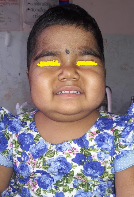
Vitals were stable, RR 28 /mt .HR 88/mt ,circulatory status stable BP 100/70, Fully conscious oriented .Apart from the pigmentation and few healed scars there were no other types of skin rashes.( Girl of  that age skin manifestations of connective tissue disorder was thought of ,Another possibility of heavy metal poisoning as the child was on long duration treatment from another system ).No cutaneous bleeding manifestations . Few points relevant about the pigmentation,Look at the palms.The creases are not pigmented .Any generalized pigmentation  in a child with weight loss possibility of adrenal pathology to be considered. In a patient who is ill nourished and prolonged fever occasionally tuberculosis can present with adrenal involvement . Pigmentation in Addison usually involves creases, scars and nipples. Look at creases and nipples, they are not pigmented   . One more Diagnostic possibility in a  pale child with pigmentation is another  important treatable condition Vitamin B12 (Even though it is not a common entity in pediatric age group.) Look at the hands and face . In B12 deficiency the knuckles and peri oral pigmentation will be prominent; here those areas are not specifically involved. The tongue also is important in B12 usually glossitis will be prominent. In this case tongue was normal .Lips was pale and angular stomatitis, right side.
DC.jpg
Apart from pallor and jaundice, no evidence of vitamin A deficiency, KF ring in the eyes 
There was pallor, jaundice, few cervical lymph nodes were present 1 cm, non tender discrete. No pedal edema, no facial puffiness. Joints were normal.
System examination GIT
Upper GI . Mouth, tongue, oral mucosa, tonsils normal
P/A.
Liver 3 cm, soft consistency. Spleen enlarged 3 cm soft. No other mass, no free fluid.
Other systems normal 
In this context two important CNS findings to be elicited .a. Romberg sign , vibration sense ,and ankle jerk  (No need for explanation)
Discussion at this stage before we proceed with investigation 
Jaundice case with urine discoloration (from history of mother). Most important possibility is bilirubin coming out through urine in a case of obstructive jaundice ( i mean there is obstruction to the conjugated bilirubin excretion, either inside liver or outside liver. ) , rarely the color may be something else. eg intravascular hemolyis eg black water fever in malaria, G6PD deficiency etc , So it is important to answer the question right. And ensure that urine is yellow itself, Not red or dark . Some occasions a condition causing liver and kidney involvement also can lead to jaundice and hematuria , eg leptospirosis  .Here no confusion urine was yellow initially , but it disappeared by the time patient reached hospital . Here stool color was normal, no other evidence of itching .Patient appeared pale .So here we are not sure , whether it was a hemolytic jaundice and the mothers history of discoloration of urine is wrong. So we consider both possibilities. 1. In view of pallor and spleen possibility of hemolysis is high 2. Liver disease responsible for jaundice and recovering, the urine became clear earlier and eyes are yet to become normal. But in the second situation, How can you explain the pallor, How can you explain the spleen. 
You may explain,,,” pallor was there earlier due to nutritional anemia, unrelated. Spleen can occur in viral hepatitis or any other infection as part of infection, not due to hemolysis . But First possibility seems more likely.
So pallor, spleen and fever of more than one week, most important entity is malaria. Then other entities with hemolysis triggered with infection eg auto immune hemolysis ,G6PD deficiency triggered by drugs..Out of this auto immune hemolysis usually the course will be much faster hemolysis and progression (not a rule), and G6PD is less likely in a female .
INVESTIGATIONS
DC1
What are the main points here? 
Urine bile salt bile pigment nil 
Predominant unconjugated bilirubin .
Look at the enzymes, all normal, Coagulation profile normal, protein level normal, So liver cell function is normal and there is no cell injury, no canalicular obstruction also. So liver enlarged must be part of congestion due to anemia, or infiltration by cells. 
Low hemoglobin 
Total leucocytes are low. Peripheral smear showed  macrocytes. Is it megaloblastic anemia? If so  how to explain this long duration of high grade fever ,unless there is another underlying reason .(eg TB intestine causing megaloblastic picture , a rare possibility , just a postulation , No bowel disturbance here ).. 
This high ESR is it due to anemia only. 
Is there a possibility of malignancy, misleading us , Occasionally non Hodgkin lymphoma may present this way .
This high ESR with a low count, and hemolysis is there possibility of SLE 
We did nt get time to answer many questions. 
When we suggested Bone marrow ,they got discharged against medical advice and got admitted in another medical college hospital . 
Investigations done from second center 
anu4
anu 2
Bone marrow done from there
anu 1
So many of the features from peripheral smear and marrow are consistent with megaloblastic anaemia .Thrombocytosis may be there, element of haemolysis may be there But we expected shift to right and this low count is a bit unusual. Another entity which comes as Differential of megaloblastic bone marrow picture is dys erythropoetic anemia .In that case shift to left ,gigantoblasts etc .Haemolysis is more likely in dys erythropoetic anaemia .One peculiarity is reticulocyte  count won’t be that high , just mild to moderate elevation .In this case corrected reticulocyte  count was 4.
 Vitamin B12 estimation done was normal .ANA negative.
There a course of vitamin B12, and other hemopoetic vitamins and micro nutrients were given. Course of antibiotics tried. Patients fever persisted after three weeks.
Blood hemoglobin remained low, in spite of three packed cell transfusions, platelets was low around 60000, but no bleeding manifestations. Total counts most of the time were on lower side and ESR most of the values were high 
Again few points controversial, why this low count? Is it part of viral infection, then why megaloblastic changes. are they unrelated entities ?.If the patient is having nutritional, megaloblastic anemia Suppose the megaloblastic picture is due to dyserythropoetic anemia (different varieties are there) can it explain the high grade fever?
He was taken to another higher center medical college.
 vitamin B12 estimation done, was normal .ANA negative .
There a course of vitamin B12, and other hemopoetic vitamins and micro nutrients were given . Course of antibiotics tried. Patients fever persisted after three weeks.
Blood hemoglobin remained low, in spite of three packed cell transfusions , platelets was low around 60000 ,but no bleeding manifestations. Total counts most of the time were on lower side and ESR most of the values were high 
Again few points controversial, why this low count? Is it part of viral infection, then why megaloblastic changes. Are they unrelated entities?.If the patient is having nutritional, megaloblastic anemia Suppose the megaloblastic picture is due to dyserythropoetic anemia (different varieties are there) can it explain the high grade fever?
He was taken to another higher center

 Evaluation at third Center 

anu10
anu8

anu11.jpg
12
ANA was repeated from there , negative
DCT  came positive once , repeat test negative
Ceruloplasmin normal, Thyroid function test normal ,Xray chest normal ,mantaux test negative. HIV negative 
Cold agglutinin negative
FNAC from Lymph node showed reactive hyperplasia
Genetic tests for chromosome breakage analysis sent . results awaited 
Patient had packed cell transfusions, A course of chlroquine , course of Vancomycin and piptaz for ten days 
Fever persisted. Pallor progressing 
Patient now re admitted here 
At the time of examination . 
Pallor , Jaundice, pigmentation all same . Spleen size 8 cm ,Liver size 3 cm , soft .
Patient in congestive failure (JVP raised , hyperdynamic precordium cardiomegaly .Liver enlargement more likely to be due to congestive failure .
No free fluid .Chest clear .
We dont have diagnosis. What are the likely possibilities in this context?
In short ……..Previously normal girl up to 9 years , having fever , jaundice and pallor and a moderate splenomegaly
Even though mother says there was urinary discoloration , most of the points argue against the jaundice due to hepatic or biliary obstruction . Four months of jaundice, no evidence of liver dysfunction . Investigations all argue against functional abnormality or cellular injury . So in view of pallor, jaundice and spleen clinically there is ongoing hemoysis. The blood parameters , corrected retic count ,high LDH and a bone marrow increased erythropoises ( focal megaloblastic ) argue for this .
As we consider the possibilities of prolonged fever of four months , at this age chronic infections, connective tissue disorders and malignancies . we need not consider hyper catablolic state , endocrinological condition etc in this case. Drug fever, autonomic disturbances seems theoretical only. So out of the major three …….which is most likely.
Malignancy, with hemolysis and jaundice unusual, still possible with NHL , but by four months at lease some clue clinically or blood investigation, So this is the last possibility .
Connective tissue disorders SLE, and one more possibility ,infection associated hemophagocytosis , possible , But many points against . 1. By this four months picture ll be more clear. Investigations even though the all the counts are on the lower side persistently , the ESR remains high , ferritin is only in hundreds. Triglycerides not high . Of course bone marrow did nt show hemophagocytosis .
So what remains is chronic infection it self
What are the possibilities   Mainly three
  1. Tuberculosis
  2. Malaria
  3. Leishmaniasis Possibility of most of the common infections like salmonella, brucella, ricketsiels , leptospirosis need not be considered in this context. HIV ruled out by investigations
Points for and against Tuberculosis…..No contact, No lymph nodes , no respiratory complaints , chest x-ray normal , mantaux negative . Pallor and low counts, bone marrow involvement may occur in TB, but in this case pallor is due to hemolysis .
Even though tuberculosis so common, it is not high in the list.
Leishmania ? Rare one, Still there are many points for leishmaniasis .Prolonged fever, weight loss, splenomegaly anemia, low counts. Pigmentation? But how common is hemolysis and frequent transfusion requirement . One of the explanation for thrombocytopenia and low count due to hypersplenism , still hemolysis seems  a bit unusual
In this context should we consider a storage disorder goucher adult type? Usually presents much earlier.
What is against malaria?
Peripheral smear never showed malaria.
No mention about rapid diagnostic test in the above tests. i don’t know whether i did not notice .
Patient had a course of chloroquine.
Are the above arguments strong enough to rule out malaria in this case?
i feel not . A patient with prolonged fever, progressively increasing spleen size, pallor requiring blood transfusions many times, malaria is top in the list. May be organism is escaping our eyes.
(We are awaiting the BM reports . we have discussed with our pathologists . to look for
a. LD bodies b.hemophagocytosis c.tuberculosis d gaucher cells , e.malaria f remote possibility of malignancy .
i have just discussed the case . Case is under my colleague. i ll let you know the further progress in coming days . 
For the preparation of this case discussion i must thank post graduates of my institution especially Anu peter and post graduates of two more Teaching institutions. Their case report and discharge summary help me to prepare this without much of typing. I don’t know who prepared these notes. Thank you my friends )

Swelling- Back of Chest

Case history ( whatever discussed and my impression i ll share for now)
One year old boy , previously had low grade fever followed by not  moving right lower limb.Minimal pain on moving upper limb also . immunized update , no history of fall .No past history of any bleeding manifestations during neonatal period and during immunizations. At the time of admission yesterday evening he was not sick .Not moving left lower limb,with main finding of restriction of hip joint movement .Other joints were normal , No focus of abscess , rashes or cutaneous bleeds. No signs of trauma .Fully conscious oriented .not allowing examination .Tone and deep tendon reflexes were normal .After sending blood for routine exam and culture patient was put on injection cloxacillin 200 mg /kg per day  and cefotaxime .Today morning baby was irritable and after noon fever became high grade and he developed the swelling back. Fast breathing proportionate to the fever , no grunt , chest air entry was normal , scattered crepitations left side .
Swelling on the back , skin was normal ,no discoloration  no signs of inflammation, tender ,no fluctuation , No crepitation elicited on touching the swelling or near areas .
staph
Blood count was High with neutrophilia .
staph .xray
Not a good xray , rotated view ,not inspiratory under penetrated. But most important is costophrenic angles both sides seen clear , No evidence of pneumothorax.There is opacity on the left side mid and upper zone. No breaking down lesions. Soft tissue does nt show any air shadows
Liver enlarged and splenic shadow obvious in X ray
Possibilities considered at this age were 
Staphylococal pneumonia with pneumothorax and surgical emphysema as the first possibility . Yes we are justified in thinking that way as a sick child of that age with fever, painful hip movement ,next day with lung signs . But air entry equal , no mediastinal shift clinically ,swelling does nt feel like surgical emphysema and xray does nt show evidence of pneumothorax 
Other DDs , is it hematoma , battered baby syndrome ? any possibility of bites and stings responsible for swelling . 
In this context pneumothorax is not there , surgical emphysema not visible ,then what is the reason for swelling ? is it abscess. Possible 
As the child is sick , we planned to make it CP and taxime and Vancomycin .
Shall update tomorrow 
18/04
Baby is sick ,toxic. Pyoderma on scalp and legs .Swelling on the back is tender ,extended upwards behind neck .Skin over swelling is now shiny and red .Not moving left limb.Irritable ,deep tendon  reflexes normal .
USG hip done did nt show collection.
Inj .CP &Vanco continuing 
Other discussions and arguments .
Why stop Cloxacillin early and step up to vancomycin ? Yes partly there is logic for this argument . We all consider Vancomycin superior to Clox in staph infections . It is not .Vancomycin has superiority only in MRSA , otherwise spectrum is small , and clox ll work better in sensitive strains. Then one more argument . Why CP ? CP to have better gram positive cover.  Cloxacillin and vanco both have very narrow range. CP have good coverage including anaerobic , but not gram negative spectrum . Then another question , why should you give third generation cephalosporins ? Right ..Here almost sure that we are dealing with an infection , and staph or gram positive organism is most likely . Here we are yet to get the culture proof. The child is ill nourished , we have send the test for HIV. In a sick child we cant take risk without giving gram negative spectrum 

A Case of Fever and Rash

7 year old girl who was healthy except for occasional episodes of wheezing presenting with fever and maculo papular rash all over body on 6th day of fever.She used to have episodes of wheezing from three year onward  which used to subside with inhalation of beta agonist and last for one or two days .No continuous medications .Last month she was hospitalized for an episode of wheezing running nose and fever ,needed hospitalization for a week . She was treated with par-enteral antibiotics .Fever and dyspnoea was relieved but morning cough was persisting . She was afebrile for a week and fever started again . This time there was  no running nose. no sore throat ,no bowel or urinary disturbances. Cough was persisting as such in the morning hours . She had some body ache , left shoulder and left elbow pain a bit more severe , but no swelling , No other joints like small joints , spine or neck .No head ache altered sensorium vomiting .In the hospital this time she was on inj. Ampicillin and Inj Cefotaxime . She was not sick , was walking around .Skin rashes appeared first on the abdomen and within 12 hours it spread through out body . Palms , soles not involved , oral mucosa genitals were not involved. No significant eye congestion ,visual problems or eye pain . No other type of skin lesions , no bleeding manifestations on skin ,subcutaneous tissue bowel or urine.There was not much of itching  , she was already on Atarax .
O/E.
Her vitals stable , (BP 100/66) ,pulse 92 per minute , good volume ,capillary refill time 2 sec . Minimal flushing of skin .Respiratory rate was 30 per minute. Her temperature 101 deg F. Fully conscious, oriented answering questions. No jaundice, no bleeds on conjunctiva , but mild congestion of palpebral conjunctiva. Cervical lymph nodes on the right side were palpable significant 1.5 cm, multiple non matted, with minimal tenderness. Few axillary lymph nodes also, same size discrete .Her anthropometric measurements showed above 80 percent weight for age, height normal. Rashes were mainly distributed around the trunk, neck and thighs especially over front aspect. Face lower legs and hands affected, but less. No lesions on palms and soles, lips, oral mucosa spared .Throat normal, No patch or bleed or ulcers in the throat, palate or oral mucosa .Joint examination showed left shoulder and left elbow pain on movement but no obvious swelling or signs of inflammation, All other joints were normal.
darshana
darshana 1
darshana 2
darshana 3
darshana 5
darshana8

Diagnostic approach.
Main features are fever and rash in  a seven year old girl child .Main group of entities which is likely to lead to this combination are
  1. Infections
  2. Connective tissue disorders
3 Fever and rash unrelated eg drug or another reason 
How ll you proceed to make out which is which 
First step 1. What about vital signs. is the child stable. There are situations where this sort of situations child is brought in an unstable state ,eg shock , respiratory distress , coma , seizure etc eg many among the above group , dengue ,meningococcemia, ricketsiels with some of the complications, connective tissue disorders with some complications , leptospirosis with encephalopathies. List is long in this situations also. You may add to this list . Few situations may be unrelated to the basic problem eg fluid loss and shock, or drug related complications. In this girl her vitals are stable .
There is another point which is helpful in the vitals. We considered the low BP state, what if the BP is on higher side? Yes it can occur, in that state possibility of connective tissue disorder, rare possibility of an infection involving kidney or causing raised ICT, drug related 
Step II
Details of fever and the relation of the rash with the fever are useful here. You may refer to the onset of rash in relation with fever. Day 1 rash , day 2 rash ,day 3 rash ,day 4 rash all argues for certain entities, They are not hard and fast rules but helpful .
Here it was high grade fever, rash appeared after five days of fever. How this is helpful? First entity any doctor in our country should consider in a patient with fever and maculopapular rash is measles, as that is the commonest entity. But is it fitting with measles? What are the strongest argument points against measles?
  1. There is no running nose.
  2. Cough not predominant, she is having cough only in the morning hours which was of same severity as she had during the asthma episode
3 .The rash did nt proceed in the usual pattern, from face, down .
4 The rash did not involve the palms, mucosa , eyes 
5 She is immunized, but mind you this point is not a strong point as we do see a lot of failure in case of single dose of measles .They come in a modified style 
  1. One point i forgot to mention in the examination, there was no Koplik spot. Koplik spot again should be used cautiously. The presence of koplik argues for measles, but absence does no t rule out. It is transient.
The time relation of rash with fever in fact is not fitting with most of the common infections. What about Rubella? Again the rash won’t be that late. The fever won’t be that high grade. Joint pain is arguing for it. But most important finding is sub occipital lymph node. Here sub occipital lymph nodes were not enlarged. One more point, useful is vaccination history. To be frank i forgot to ask this point. i ll ..She is in the ward. 
What about other viral infection?
Her vitals stable,
Does that totally rule out dengue?  . No it is still possible. Only severe grades of dengue need have unstable vitals .One point against is the late onset of rash, a bit unlikely. Joint involvement is arguing for chickun guinea, Absence of muscle tenderness, conjunctival congestion is arguing against. Still we need to consider dengue and chicken guineas as strong possibilities. So we were looking for other signs like cutaneous bleeds, Of course here before we send for lab investigations we can do Tourniquet test on the bedside. (To be frank we did not do it. Only when i write this i remember what all things we are missing when we actually do in the ward) 
Few more of the viral infections which can present with fever, like parvo , adeno , coxakie ,echo etc but a high grade fever and fever lasting this long we should not consider this entities as we may miss important entities 
What other infection, treatable, which we did nt consider so far, can present with fever and maculopapular rash, without running nose? Leptospirosis . Here what points for and against? In fact many points are not arguing for. No muscle pain, No jaundice, No bleeding tendency, P/A no liver spleen. There are few lymph nodes in the neck, minimal conj congestion .Can we rule out that possibility totally? NO, being a treatable entity, and likely to have modified manifestation this entity should be one of the DDs always, till you rule out for sure. And empirical treatment is justified if you are not in a position to rule out 
Should we consider other bacterial infections which can cause fever eg enteric fever .Enteric fever can cause rash, but not this extensive and lasting. So less chance for enteric fever  directly leading to this rash. Of course we were considering the possibility of drug rash on another condition with fever. She was on cefotaxime and Ampicillin for two days before she was referred here. Does that remind you something which you learned? In a case of fever, rash. And relation of Ampicillin. Yes Infectious mononucleosis. Of course that possibility to be considered high in the list. Fever, rash , few lymph nodes in the neck , axilla. Hepato splenomegaly need not be there in all cases. Throat was normal , this is a better point to argue against IMN , but again not in hundred percent of case. Absence of jaundice, you may use for argument, Again it is not a hard point.  Toxo plasma and cytomegalovirus causes similar picture which the investigation only will rule out 
Which other infections we did nt consider so far?
Ricketsiels, secondary syphilis. Lot of atypical presentations of ricketsiels we encounter now in this area. The rash typically involves the palms and soles. And we should look for Eschar. in this case both were not there. 
So next thing. 
Are we dealing with infection? is it something else 
Is it drug ?
is it connective tissue disorder ?
Drug as a reason can t be rule out for sure, as this occurred on the second day of antibiotics. Not much of itching. no mucosal involvement are weak points to argue against that possibility . So we stopped all drugs. In this case we don’t have a firm diagnosis. Patient’s vitals stable.
Should we consider possibility of connective tissue disorder? Yes , we should , in a girl child of this age. They can present like this. May be acute. But of course common things common , we consider infection possibility as 90 percent , and the investigations for Connective tissue disorders are costly ,she did nt have any major markers to argue for connective tissue disorder. So we planned to do the costly investigations in the next stage. Of course we ll do the basic minimum in all cases eg counts. ESR, CRP, Urine which may give some clues in this line 
Now the investigations 
Total count 8000 /cmm. i dont remember the HB i think it was normal
DC Neutrophil 70 %,Lymph25 E 5. Platelet 2.7 lakh
ESR 70 
Urine alb  nil, microscopy nil , no bile salt ,bile pig
Renal function Urea 18, Creatinin 0.9
LFT , bilirubin 0.9 total and direct 0.2 ,Total proteiin 7 , alb 4 , SGPT  45,SGOT 40 
So where are we now? 
Counts, not that high, with a high ESR. Normal Urine parameters and Normal LFT. i can defintely rule out Lepto . Only High ESR is arguing all other parameters against .Even anicteric lepto SGPT ll be high 
Count does that suggest dengue. Not that low, normal platelets. and high ESR . Not suggestive, but can’t rule out. So we send for Ig M dengue. Not NS 1 as it is already 7th day
What about IMN , Toxoplasma, Cytomegalovirus. Yes possibility is unlikely with this count and neutrophil predominant picture. Still we send for a peripheral smear. One more dictum, any prolonged fever peripheral smear thick and thin should be done as we are living in India and malaria can be one of the DDs of any of this sort of cases. I am not sure what was the result of PS. i ll update tomorrow 
So what about possibility of a connective tissue disorder 
Should we consider Kawasaki ?
This age, very rare , No mucosal involvement , chances are very low
High ESR with Low count, always SLE to be ruled out 
So we plan to rule out in the next week.
What did we do? 
My first diagnosis in this case of fever with rash happening on the seventh day, not sick with few lymph nodes , normal system findings was ricketsiels , atypical manifestation 
Yesterday we send for Weil Felix and IgM Eliza for Scrub typhus 
As we were not sure of drug as a reason, we took all investigations , stopped all parenteral antibiotics and put her on Doxycycline 100 mg daily 

temp chart of darshana
Today morning she is afebrile 
We are yet to get the report of tests . I ll update 
19/04/2016
Weil felix test came as negative 
IgM Eliza not done
So where are we  now .
As jom suggested , may be it is just viral infection with rash spontaneously remitted and we were attributing response to doxicycline
Weil felix may be false negative 

Nephrotic syndrome

Three year old girl only child born to a  young couple was diagnosed as Nephrotic syndrome at one and half years of age. No consangu...