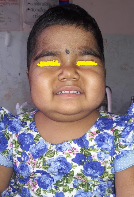
( This case was admitted in November 2012 pediatric department under my colleague Dr.T.D.Muraleedharan s Unit just for a day . They went against medical advice the very next day and got admitted in two more other medical colleges. After a gap of three months they are back again in our institution now , under Dr. Muraleedharan . As it is an interesting problem sharing my thoughts )
Eight and half year old girl child from poor social back ground presented with fever , pallor and jaundice four months Third child born out of non consanguineous marriage . She was ill nourished throughout but never had any significant illness requiring hospitalization .In November she had low grade fever followed by small vesicles on the limbs and face which was not itching , not painful .There were no mucosal lesions. No drugs before the eruption .They lasted four days followed by healing with scarring .Fever persisted .Two weeks later mother noticed yellowish discoloration of urine and eyes. Stool normally colored, not pale no blood stain or malena .No rashes or itching associated with this .Sleep normal, no alteration of sensorium. Continued ayurvedic treatment .As the patients fever persisted she got admitted in our institution in December 2015. The fever was intermittent, high grade occurring daily, no diurnal variation. No chills or rigor .Mother noticed gradual black staining of skin of face trunk and skin .Apart from the fever there was no bowel disturbances, no difficulty in micturition.Acording to mother urine color gradually cleared but the eye color persisting. No history of abdominal pain, joint pain weakness. She was able to do her day to day activities without help.


Vitals were stable, RR 28 /mt .HR 88/mt ,circulatory status stable BP 100/70, Fully conscious oriented .Apart from the pigmentation and few healed scars there were no other types of skin rashes.( Girl of that age skin manifestations of connective tissue disorder was thought of ,Another possibility of heavy metal poisoning as the child was on long duration treatment from another system ).No cutaneous bleeding manifestations . Few points relevant about the pigmentation,Look at the palms.The creases are not pigmented .Any generalized pigmentation in a child with weight loss possibility of adrenal pathology to be considered. In a patient who is ill nourished and prolonged fever occasionally tuberculosis can present with adrenal involvement . Pigmentation in Addison usually involves creases, scars and nipples. Look at creases and nipples, they are not pigmented . One more Diagnostic possibility in a pale child with pigmentation is another important treatable condition Vitamin B12 (Even though it is not a common entity in pediatric age group.) Look at the hands and face . In B12 deficiency the knuckles and peri oral pigmentation will be prominent; here those areas are not specifically involved. The tongue also is important in B12 usually glossitis will be prominent. In this case tongue was normal .Lips was pale and angular stomatitis, right side.

Apart from pallor and jaundice, no evidence of vitamin A deficiency, KF ring in the eyes
There was pallor, jaundice, few cervical lymph nodes were present 1 cm, non tender discrete. No pedal edema, no facial puffiness. Joints were normal.
System examination GIT
Upper GI . Mouth, tongue, oral mucosa, tonsils normal
P/A.
Liver 3 cm, soft consistency. Spleen enlarged 3 cm soft. No other mass, no free fluid.
Other systems normal
In this context two important CNS findings to be elicited .a. Romberg sign , vibration sense ,and ankle jerk (No need for explanation)
Discussion at this stage before we proceed with investigation
Jaundice case with urine discoloration (from history of mother). Most important possibility is bilirubin coming out through urine in a case of obstructive jaundice ( i mean there is obstruction to the conjugated bilirubin excretion, either inside liver or outside liver. ) , rarely the color may be something else. eg intravascular hemolyis eg black water fever in malaria, G6PD deficiency etc , So it is important to answer the question right. And ensure that urine is yellow itself, Not red or dark . Some occasions a condition causing liver and kidney involvement also can lead to jaundice and hematuria , eg leptospirosis .Here no confusion urine was yellow initially , but it disappeared by the time patient reached hospital . Here stool color was normal, no other evidence of itching .Patient appeared pale .So here we are not sure , whether it was a hemolytic jaundice and the mothers history of discoloration of urine is wrong. So we consider both possibilities. 1. In view of pallor and spleen possibility of hemolysis is high 2. Liver disease responsible for jaundice and recovering, the urine became clear earlier and eyes are yet to become normal. But in the second situation, How can you explain the pallor, How can you explain the spleen.
You may explain,,,” pallor was there earlier due to nutritional anemia, unrelated. Spleen can occur in viral hepatitis or any other infection as part of infection, not due to hemolysis . But First possibility seems more likely.
So pallor, spleen and fever of more than one week, most important entity is malaria. Then other entities with hemolysis triggered with infection eg auto immune hemolysis ,G6PD deficiency triggered by drugs..Out of this auto immune hemolysis usually the course will be much faster hemolysis and progression (not a rule), and G6PD is less likely in a female .
INVESTIGATIONS

What are the main points here?
Urine bile salt bile pigment nil
Predominant unconjugated bilirubin .
Look at the enzymes, all normal, Coagulation profile normal, protein level normal, So liver cell function is normal and there is no cell injury, no canalicular obstruction also. So liver enlarged must be part of congestion due to anemia, or infiltration by cells.
Low hemoglobin
Total leucocytes are low. Peripheral smear showed macrocytes. Is it megaloblastic anemia? If so how to explain this long duration of high grade fever ,unless there is another underlying reason .(eg TB intestine causing megaloblastic picture , a rare possibility , just a postulation , No bowel disturbance here )..
This high ESR is it due to anemia only.
Is there a possibility of malignancy, misleading us , Occasionally non Hodgkin lymphoma may present this way .
This high ESR with a low count, and hemolysis is there possibility of SLE
We did nt get time to answer many questions.
When we suggested Bone marrow ,they got discharged against medical advice and got admitted in another medical college hospital .
Investigations done from second center


Bone marrow done from there

So many of the features from peripheral smear and marrow are consistent with megaloblastic anaemia .Thrombocytosis may be there, element of haemolysis may be there But we expected shift to right and this low count is a bit unusual. Another entity which comes as Differential of megaloblastic bone marrow picture is dys erythropoetic anemia .In that case shift to left ,gigantoblasts etc .Haemolysis is more likely in dys erythropoetic anaemia .One peculiarity is reticulocyte count won’t be that high , just mild to moderate elevation .In this case corrected reticulocyte count was 4.
Vitamin B12 estimation done was normal .ANA negative.
There a course of vitamin B12, and other hemopoetic vitamins and micro nutrients were given. Course of antibiotics tried. Patients fever persisted after three weeks.
Blood hemoglobin remained low, in spite of three packed cell transfusions, platelets was low around 60000, but no bleeding manifestations. Total counts most of the time were on lower side and ESR most of the values were high
Again few points controversial, why this low count? Is it part of viral infection, then why megaloblastic changes. are they unrelated entities ?.If the patient is having nutritional, megaloblastic anemia Suppose the megaloblastic picture is due to dyserythropoetic anemia (different varieties are there) can it explain the high grade fever?
He was taken to another higher center medical college.
vitamin B12 estimation done, was normal .ANA negative .
There a course of vitamin B12, and other hemopoetic vitamins and micro nutrients were given . Course of antibiotics tried. Patients fever persisted after three weeks.
Blood hemoglobin remained low, in spite of three packed cell transfusions , platelets was low around 60000 ,but no bleeding manifestations. Total counts most of the time were on lower side and ESR most of the values were high
Again few points controversial, why this low count? Is it part of viral infection, then why megaloblastic changes. Are they unrelated entities?.If the patient is having nutritional, megaloblastic anemia Suppose the megaloblastic picture is due to dyserythropoetic anemia (different varieties are there) can it explain the high grade fever?
He was taken to another higher center
Evaluation at third Center


ANA was repeated from there , negative
DCT came positive once , repeat test negative
Ceruloplasmin normal, Thyroid function test normal ,Xray chest normal ,mantaux test negative. HIV negative
Cold agglutinin negative
FNAC from Lymph node showed reactive hyperplasia
Genetic tests for chromosome breakage analysis sent . results awaited
Patient had packed cell transfusions, A course of chlroquine , course of Vancomycin and piptaz for ten days
Fever persisted. Pallor progressing
Patient now re admitted here
At the time of examination .
Pallor , Jaundice, pigmentation all same . Spleen size 8 cm ,Liver size 3 cm , soft .
Patient in congestive failure (JVP raised , hyperdynamic precordium cardiomegaly .Liver enlargement more likely to be due to congestive failure .
No free fluid .Chest clear .
We dont have diagnosis. What are the likely possibilities in this context?
In short ……..Previously normal girl up to 9 years , having fever , jaundice and pallor and a moderate splenomegaly
Even though mother says there was urinary discoloration , most of the points argue against the jaundice due to hepatic or biliary obstruction . Four months of jaundice, no evidence of liver dysfunction . Investigations all argue against functional abnormality or cellular injury . So in view of pallor, jaundice and spleen clinically there is ongoing hemoysis. The blood parameters , corrected retic count ,high LDH and a bone marrow increased erythropoises ( focal megaloblastic ) argue for this .
As we consider the possibilities of prolonged fever of four months , at this age chronic infections, connective tissue disorders and malignancies . we need not consider hyper catablolic state , endocrinological condition etc in this case. Drug fever, autonomic disturbances seems theoretical only. So out of the major three …….which is most likely.
Malignancy, with hemolysis and jaundice unusual, still possible with NHL , but by four months at lease some clue clinically or blood investigation, So this is the last possibility .
Connective tissue disorders SLE, and one more possibility ,infection associated hemophagocytosis , possible , But many points against . 1. By this four months picture ll be more clear. Investigations even though the all the counts are on the lower side persistently , the ESR remains high , ferritin is only in hundreds. Triglycerides not high . Of course bone marrow did nt show hemophagocytosis .
So what remains is chronic infection it self
What are the possibilities Mainly three
- Tuberculosis
- Malaria
- Leishmaniasis Possibility of most of the common infections like salmonella, brucella, ricketsiels , leptospirosis need not be considered in this context. HIV ruled out by investigations
Points for and against Tuberculosis…..No contact, No lymph nodes , no respiratory complaints , chest x-ray normal , mantaux negative . Pallor and low counts, bone marrow involvement may occur in TB, but in this case pallor is due to hemolysis .
Even though tuberculosis so common, it is not high in the list.
Leishmania ? Rare one, Still there are many points for leishmaniasis .Prolonged fever, weight loss, splenomegaly anemia, low counts. Pigmentation? But how common is hemolysis and frequent transfusion requirement . One of the explanation for thrombocytopenia and low count due to hypersplenism , still hemolysis seems a bit unusual
In this context should we consider a storage disorder goucher adult type? Usually presents much earlier.
What is against malaria?
Peripheral smear never showed malaria.
No mention about rapid diagnostic test in the above tests. i don’t know whether i did not notice .
Patient had a course of chloroquine.
Are the above arguments strong enough to rule out malaria in this case?
i feel not . A patient with prolonged fever, progressively increasing spleen size, pallor requiring blood transfusions many times, malaria is top in the list. May be organism is escaping our eyes.
(We are awaiting the BM reports . we have discussed with our pathologists . to look for
a. LD bodies b.hemophagocytosis c.tuberculosis d gaucher cells , e.malaria f remote possibility of malignancy .
i have just discussed the case . Case is under my colleague. i ll let you know the further progress in coming days .
For the preparation of this case discussion i must thank post graduates of my institution especially Anu peter and post graduates of two more Teaching institutions. Their case report and discharge summary help me to prepare this without much of typing. I don’t know who prepared these notes. Thank you my friends )


No comments:
Post a Comment