Friday, 2 December 2016

Case of empaema , simple problems during management

Four year old girl child presented with fever cough and dyspnea. No running nose ,cough was dry . 
She was hospitalized and was on Inj Ceftriaxone. 
As the dyspnea worsened she was referred to us.
First child born at term out of uneventful antenatal history was not asphyxiated. She had neonatal convulsions and was hospitalised for twenty days ? meningitis. 
Development was normal .
Last year she developed thrombocytopenia ,diagnosed and managed as ITP . 
There is history of abscess drained three times from different sites. Immunized update. 
No family history of any significant illness. ( No consanguinity) . No contact with tuberculosis. 
No past history of recurrent respiratory infection,ear discharge,purulent nasal discharge ,loose stools .

On admission she was dyspneic, fully-conscious, maintaining saturation above 90 percent with 50 percent oxygen circulatory status stable .
No focus of pyoderma or abscess. 
No rash or bleeds,
No Lymph nodes 
 No stigmata which grossly suggest inherited immunodeficiency . 

Her weight , height and Mid arm circumference was adequate for her age .
Respiratory system exam 

Upper airway normal 

Chest movement diminished on the right side.
 Apex and trachea shifted to left. Percussion stony dull in the right lower inter scapular axillary infra mammary areas. Air entry diminished same areas VR decreased ,no bronchial breathing, no added sounds. 
CVS normal except shift of apex to left 
P/A no organomegaly, no free fluid 
CNS fully conscious, no neurological deficit no Signs of meningeal irritation 

In short 

Four year old child admitted with fever ,dry cough and progressively worsening dyspnea. 
 Normal upper airway and mediastinal shift brings pathology below tracheal bifurcation. Mediastinal shift to left and stony dullness argue collection inside right pleural cavity. In view of fever in this age group most common reason is bacterial pneumonia with syn-pneumonic effusion. But here the cough is dry only and more  features of fluid in pleural cavity than parenchymal involvement. Possibility of empaema considered high . 

Is there an immunodeficienty behind ? 

Teaching points for PGs at this stage of history .


Multiple abscesses drained ,and now  empaema possibility of susceptibility to staph infection high ? Yes we should investigate in that line. Immunodeficiency states with abscess formation are 
1. Problems with phagocytosis of neutrophils 
2. Problems with respiratory burst ,chronic granulomatous disease. Usually boys but girls may be affected 
3. Jobs syndrome ,
other entities also 
Neutrophil related problem and bacterial infection . It may be adhesion problems , chemotaxis , phagocytosis or respiratory burst . 
In problems with adhesion , and chemotaxis, neutrophils wont reach the site of organism . So NO evidence of inflamation, no pus . Common reason for this state is any significant neurtopenia. ( No neutrophils to reach the site ) . In adhesion problems usually the neutrophil count in the peripheral blood ll be high ( They wont go out ,but remain inside )

Course 

USG confirmed empaema right side and ICD done .She was put on inj CP , Cloxacillin 200 mg/kg /day and Gentamicin .

Temperature touched baseline in two days .She was active ,playful .Exam showed trachea and apex in normal space ,dullness in the right infrascapular area , decreased VR and breathsounds same area.
So some more of pus remaining


Repeat Xray chest and USG was done 


On the right side costophrenic angle was clear ,patchy pneumonia middle lobe. No abscess .
On the left side there was a circular translucency . 

Two questions remained

1.What is the reason for the Clinical findings in the right infrascapular area. Xray and repeat USG did nt show fluid .So this was attributed to pleural thickening (not a good explanation as this usually it takes two weeks )

2.What is the translucent shadow on the left side.?


Possibilities for this shadow 

1. cavity inside parenchyma .
2. Pleural adhesions 
3.abdominal viscera 
4.Pericardial cyst 

Out of this possibility of abdominal viscera was less likely. ( diaphragmatic hernia and eventeration are the common ones , but here diaphragm is seen in normal place . Other possibility of rapture is less likely as there is no trauma involved. 

This location pericardial cyst unlikely ( this large cyst anterior to heart 

So main DDs considered were a partially filled abscess inside parenchyma and pleural adhesions causing an appearance like this .
If it is parenchymal it must be in the lower lobe. 
If pleural it can either be in the front or back 
The line of diaphragm could be traced medially as there was a confusion whether this represented level of fluid. One more argument against abscess was the translucency of air ( above the fluid, if it is an abscess ) was not dark enough. 
So 
Repeat X ray AP and Lateral was taken 


So Now ?


There is no shadow in the lower lobe area as we postulated. The shadow in the mid zone represent the middle lobe consolidation. About the shadow we were considering is in the anterior part , and the level is clear , traceable beyond the diaphragm . It is a partially filled abscess. 
Rechecked for clinical findings in the same area. Normal resonance in percussion ,Normal intensity of vesicular  breath sounds ,normal resonance .

She remained a febrile ,active except for the fact that pus was draining 40 to 50 ml per day . Air was bubbling episodically . 
As there is possibility of bronchopleural fistula and lung expanded well we did nt ask her to inflate balloon which we used to do .
As she remained afebrile ,not sick not dyspnoeic same antibiotics were continued .We were curious from where this air and pus was coming as the X ray and USG did nt show any collection. As there was doubt whether there is chance of air entering to the tube from outside by accidental withdrawal , we checked for that also , and ensured ICD tube  has entered enough .The air column was moving free .

Two other things which came in to consideration

1. Should we instill streptokinase ?. We used to and had good results. In this case we did nt . Because there is bronchopleural fistula and we were afraid whether this will affect the sealing of fistula . 
2.Second point . Should we go for VATS. Yes . Many institutions resorts to early VATS and good results. Here in our institution it is being done, but not many cases and not for these young ones . We discussed with parents about the option to send to higher institution , but they opted to continue treatment here 

On the tenth day of ICD the bubbling was significant . 


There was argument for and against stepping up antibiotic . Consensus was to step up. 
What were the arguments for continuing same drugs . Kid is not sick , not febrile . Lung findings were not worsening ,xray and USG shows clearance. The shadow in the left side also does not need any intervention apart from what she is getting . 
Opposite view was..Patient is  already on ten days of antibiotics. Pus still coming , broncho-pleural fistula is there. Is it safe to continue first level antibiotics ?. Yes staph is always dangerous. ( One point forgot to mention that culture did nt yield growth )

We decided to step up 

Next question , stepping up to which one. Vancomycin ? Linezolid Clindamycin ? 
Vancomycin is superior for MRSA ,but inferior to cloxacillin or other antistaph agents if not MRSA .In this case chance for  MRSA is less. She responded very well to first line . Spectrum of action of vancomycin against other organisms less . 
Next choice was between Linezolid and clindamycin. Our experience with Linezolid for the last few years were excellent ,Good response most of the kids tolerating the drug very well . 
She was put on Linezolid 10 mg per kg per dose three times daily. Today fourth day of Linezolid. The air leak stopped completely, the pus drainage reduced. No pus yesterday. We were planning to remove the tube today . But today ten ml of pus still there. Now we decided to continue ICD for few more days . 


What are our plans now

1.First control present problem 

Next step to find out the reason behind , ie there is immunological basis . We are planning to do the immunoglobulin levels. We had hyper igE syndromes presenting like this. One of those cases now going well with long term co trimoxazol orally low dose .
If results are negative, may need to consider rarer entities. 
Shall let you know the follow up 


8/8/2016
On sunday she developed sudden dyspnea. Xray was taken and showed this 

ICD was blocked ,resulting in pyopneumothorax .ICD changed and distress releived . Fifty ml of pus drained . 

Linezolid stopped and she was put on Vancomycin and cefepime . 
Next day draining of pus almost nil but the air bubbling continued, with each breath .
In view of this CT was done 


;


Broncho pleural fistula . 
ICD was continued for three weeks. Gradually bubbling stopped . We could remove ICD ,patient afebrile , about to shift her to ward 

She developed sudden dyspnoea cyanosis
 Xray taken 


Again ICD was put 

This time her dyspnea did nt improved. Bubbling continued. 
We decided to surgical intervention . Patient opted for Surgery from Another institution 

20 , december 

Middle lobectomy done . 
Specimen 


Post surgery Xray 
She is doing fine now 


Saturday, 12 November 2016

Child with abdominal distention

one and half year old female child was referred to us by her family doctor as he thought something wrong with her abdomen.

She was second kid born out of non consanguineous marriage ,elder one is five year old healthy boy .
No significant antenatal ,perinatal problems . 
Her development was normal in all fields till ten months when she started to stand holding furniture ,from there achievement was slowed down .
Now She can  walk holding on fingers which corresponding to one year. 
Her manipulation ,social development corresponds to one year and three months. 
In short there is mild delay in all fields , gross motor  a bit more involved. 
Parents took her to the family doctor when she developed running nose and fever and fever  subsided in three days with symptomatic measures. Parents did not notice anything wrong with her abdomen . They were a bit concerned with the developmental delay because the elder boy achieved all the milestones earlier than her 

No seizures ,no excessive irritability .
No history of any significant illness so far . 
No contact with tuberculosis. 
No history of travel outside .


O/E 
vitals stable .
No pallor 
No significant lymph nodes. No rashes, No bleeds .

GIT 

Oral cavity normal

Abdomen exam showed Liver 5 cm , span 10 cm firm ,sharp margins surface smooth surface
Spleen 12 cms ,crossing the mid line. Firm .Possibility of other masses ruled out as insinuation of fingers between costal margin not possible and bi manual palpation negative .
Genitals normal
Nervous system examination  
Cranial nerves all normal . Special stress on eyes. No squint ,no abnormal eye movements, Ocular movement in all directions full 
( fundus normal ,no cherry red spots or optic atrophy ) , 

Discussion

In short girl child one and half years with delay in milestones , with gross spleno hepatomegaly . 
Overall pattern of development she was normal up to nine months but there is slowing .Deciding about the development is one major decision
1.Is this delay significant or just a normal variation ?
2. Is it static type or Progressive type ? 
A bit difficult ,because we do see mild slowing of the previous pace of development following any acute illness. So may be this is just a normal situation 
But  few points need to be answered before we are happy with the above argument 
1. Was there a significant illness to account for this delay?
2. Of course  significant illness can cause some slowing or may be a dipping of the curve , and after a gap of few week catch up . But in this case it is more than three months since they noticed the dip 
3. We do get a dip or slowing following acute illness severe enough. But will it affect all the fields . Usually gross motor is the maximum affected. Here all the fields are affected . 
So in short pattern of development is more like a progressive disorder . But we dont have strong evidence for it as there is nothing suggestive of a definite grey or white matter involvement so far .


Once more a rethinking 
As the  development is affected and a gross spleen is there  bit of bias in our approach  that it is a neurological problem  . Did we jump to that conclusion without a second thought ?
Of course hard finding is significant spleno hepatomeglay in a young kid ? Other points are not strong enough. May be the basic entity responsible for the gross organomegaly cause bit of delay as part of its course eg a chronic infection .
So we ll take a deviant approach ignoring  the development part which is not a hard finding 
We ll take the dependable finding of gross spleno hepatomegaly here 
What are the main entities which cause this much of organomegaly at this age ?

1. Hemolytic anemia esp thalassemia 
2. Malaria 
3.Malignancy ,adult type of Chronic myeloid leukemia ,rarely NHL
4.Kala Azar 
5.Other hematological disorders like osteopetrosis, Myelofibrosis 

 She is not pale, so far no requirement of blood transfusion. 

There is no fever apart from short febrile illness lasting for two days which subsided

No weight loss 

These arguments argues against most of the above differentials. So this must be an entity causing significant enlargement of spleen and liver ,uniformly a bit firm in consistency ,without involving blood formation or causing blood element consumption . This is one entity not causing much of fever or inflammatory element .
Considering the above points revise the list again , excluding the above 
Storage disorder
Portal hypertension 
Portal hypertension without any features of liver function involvement and this huge spleen a bit unusual , not impossible .

 if we give significance to development problem storage disorder  is more likely.
Which one ? carbohydrate ?Lipid.? Others like Mucopolysacharide, mucolipidosis and many more . 
Here no features in the general examination supporting the last groups , 
Never patient had symptoms of hypoglycemia. But glycogen storage disorder may not have symptoms ,may present with hepatomegaly only . 
But spleen if at all is possible in Type 4 and it is due to portal hypertension ( reminding you , glycogen is not stored in spleen .Glycogen is stored in liver, muscles including heart , and kidneys Not in spleen . Spleen is involved in Type 4 not with storage but by portal hypertension ) This huge spleen is unlikely in portal hypertension .
Apart from this few of the general examination features which may be helpful when we consider storge of glycogen are the doll facies, and floppiness, large toungue etc. In this case none of them . 
So most likely not glycogen storage. 
Which one of lipid storage ? All of them can have liver and spleen enlargement . Main group are gangliosidosis 1 and 2, Spingolipidosis and cerebrosidosis 
Gangliosidosis Type I look like MPS and they ll have abnormal appearance and obvious features. Type II. Tay sachs wont cause organomegally , Sandoffs organomegaly possible but small . and onset late 
Neiman pick many verities are there  , most of them cause predominant liver involvement . Most of them significant functional disturbance esp type C . neurological involvement esp occular , gaze etc. more prominent. Of course fundus examination ll help as all  of them may have cherry red spots  . But it is not a must in early stages. 
Here none of the above descriptions fit with the clinical feature.
So what is remaining is Gauchers , adult type. 
Out of the two types. infantile type ( adult does nt mean it ll occur in older and vice versa ) will have severe neurological problems. Here except for a minimal delay neurological examination is normal 
So we considered the possibility of Gauchers 
So after the basic blood counts we decided to go ahead with a bone marrow examination 
We personally went to pathology department to discuss the problem and we wanted them to look for 
1.Leishmaniasis
2. Malarial parasite
3.Malignancy both CML , or NHL
4.Gauchers 

This is the Bone marrow picture 


( We thank Doctors in our Pathology department ,especially Dr.Feroz , HOD Pathology for giving us the slide )
Patient sent home this week 
We are planning an enzyme assay . The firm which markets the enzyme ll do the enzyme estimation free of charge. We have contacted them , and the tools for collection of sample awaited. 
One happy news .. 
Till recently cost of the enzyme replacement was around fourty lakh a year and no chance for a poor family to afford this for a lifetime . Last year one family filed a case and the verdict is favoring them , to support the cost by the govt. 
I reserve my comments about the court decision and the ethical issue involved . 
But we ll reveal the information to this family too .

Thursday, 3 November 2016

girl with neck pain


Six year old girl child presented with difficulty in turning neck for last two months.Movement of neck in all directions were painful , and was progressively increasing. 

Occasional low grade fever,which was controlled with paracetamol.
No neck  swelling or swelling elsewhere noticed by parents.
No history of trauma 
No history of bone or joint pain elsewhere .
No skin rashes or bleeds.
No past history of significant illness . 
No contact with tuberculosis. 

Immunized update


She was pale  No lymph node enlargement . 
No bleeding manifestations
 Movement of neck restricted in all directions and torticollis present 
She does nt have any swelling or pain on any othe bony points

Abdomen exam
No hepato splenomegaly 

We did a neurological examination ............Normal . 

IMAGINGS 


Xray Neck lateral view 


First look it may be passed of as normal . But few abnormalities 

Common mistakes in interpretation of Xray neck ( both lateral and PA ) 

1. Missing abnormalities in the lowest cervial vertebra. If not taken properly the shoulder shadow may mask the vertebra
2. Missing features of Axis and atlas and the relation 
Before looking at the bodies try to draw a line along anterior posterior margin of bodies. canal and the spine. Usually it ll be possible 
Here there is straightening . Inter vertebral space normal , Bodies look normal except the fourth . Axis looks denser . Ring of atlas and atlanto axial relation normal 


Seeing this X Ray , she was re examined for bone and joint problems .It showed found out small swelling on forehead and painful areas on her scalp. There was pediculosis but no infected lesions to account for the pain 


Plane Xray skull lateral view 


Erosion of skull bones multiple sites ,without discrete margins sort of moth eaten appearance..Areas of sclerosis .

MRI brain and Spine 


Look at the Skull bones ..


Brain parenchyma , Pituitary Hypothalamic area was normal 


Cervical spine . the atlas and axis are involved. Lower level multiple vertebral bodies , thoracic and lumbar involved. 

No evidence of compression on spinal cord or medulla .
Reexamination CNS was done ,All deep tendon reflexes normal ,sensations normal
Two common entities at this age

1. Neuroblatoma 
2,Langerhan cell disorder . Eosinophilic granuloma
3 Chronic recurrent multifocal osteomyelitis 


Other Investigations 


Blood , Hb 6 grams, Total count 8000, N 60 ,L38,E2

Mantauxe negative 

Peripheral smear  showed  microcytic hypochromic RBCS 

WBC series and platelet count and distribution were normal 

 VMA 24 hour urine.....was  normal

USG abdomen . Liver , spleen normal ,No Mass Adrenals normal .

Xray chest .No  rib erosion, No mass in the chest. lung and heart shadow and mediastinum were normal 

Needle aspiration cytology Not done 

Bone marrow done  ...., No evidence of leukemia , lymphoma or neuroblastoma. 

Chronic Recurrent Multifocal Osteomyelitis is a rare entity 

( CRMO usually involves long bones than skull and spine even though it is possible . Here most of the long bones spared .Examination of clavicle did nt show any swelling or tenderness . Xray showed clavicles were normal .Clavicle is one of the commonest bone involved in CRMO)

We considered  Langerhans cell disorders as first possibility 

 .

She was referred to Regional Cancer center and she is on treatment from there

Tuesday, 1 November 2016

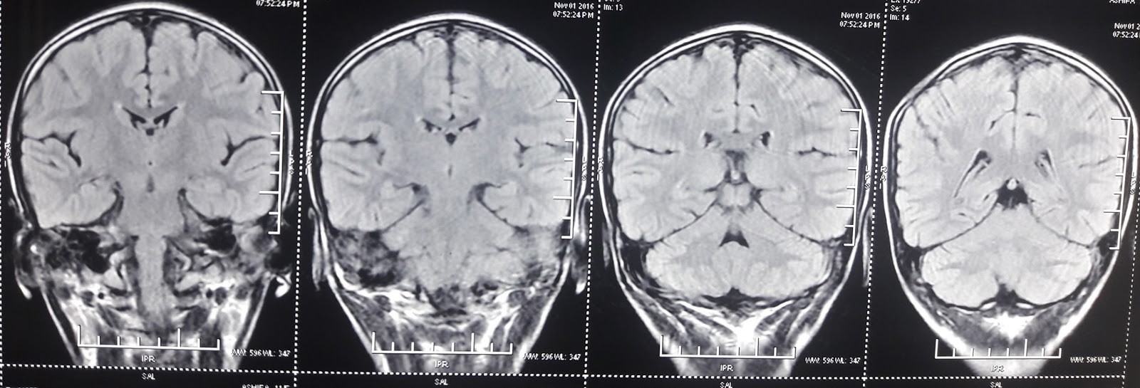
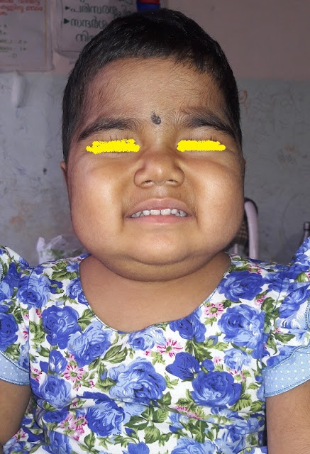
Case of Cerebral palsy

11 year old girl with developmental delay.

Born out of non consanguineous marriage without any significant family history of  seizures, mental retardation ,early childhood deaths .
No antenatal insults ,delivered at term ,cried immediately after delivery ,No neonatal problems. Initial developments were normal .Mother noticed the milestones were lagging after  sitting onward. Now her Gross motor development corresponds to 7 years .Fine motor corresponds to seven years. Her language, social normal . She can read and write both English and Malayalam She can subtract ,multiply and divide numbers .

Step wise analysis 

There is developmental delay

Delay predominantly in Gross motor and fine motor  areas. Social and language normal

Delay is of Static type.That is gradually achieving milestones ,no regression  .
Hence at this stage an entity causing developmental delay in gross motor and fine motor only with a pattern of static nature considered 

When she was one and half years parents noticed abnormal jerky movements mainly involving her upper limbs and neck during episodes of fever. Initially it was attributed to drugs given for the fever.This disappeared completely after the fever in the initial episodes. There was no alteration of sensorium during these episodes. They disappeared during sleep.
Initial years she did nt have many episodes of fever. At the age of four she was investigated in NIMHANS with imaging ,biochemical tests and diagnosed as cerebral palsy ,mixed 

She is now eleven years going to school, In the school her performance in mathematics is very good , Reading and understanding Malayalam ,English and hind . But her writing is bad . She writes her own ideas but occasional alphabets slips of from her pen tips. 
She is brought now as the mother is concerned with her day to day activities .in between the episodes of fever also  She drops things from her hands frequently . She cant drink from glass ,she spills . She cant walk holding anything straight , While trying  to walk with a cup of tea her facial expression changes grossly in the effort to keep cup with water in her hands .


Examination 

Vitals stable ,She is thin with normal height . Head circumference normal . Hair and skin normal , No dysmorphic features. No neurocutaneous markers .
No KF ring in the eyes .
On the left eye conjunctiva shows a prominent vascular engorgement on the sclera. No significant features on the other areas .No abnormal movements during rest .She  keep her neck tilted to right side most of the times.but all neck movements normal 

Nervous system examination 

Intelligence memory and speech normal 
Cranial nerves normal . Normal vision , fundus 
Motor system ,Bulk ,power normal , Spasticity bilaterally in all limbs more in the lower limbs ,no asymmetry .Deep tendon reflexes all exaggerated ,no patellar or ankle clonus. No rigidity 
( When we suspect possibility of pyramidal and extrapyramidal lesions there can be a combination of spasticity and rigidity .Spasticity after the give way feeling movement is free. If there is associated rigidity this wont be the case. 
Now her abnormal movements are not obvious , spills on drinking from cup 
Sensations all modalities normal
No cerebellar signs 
Peripheral nerves not thickened
Skull and spine normal 
Other systems normal .
At present 


doubtful telangiectasia on the conjuctiva. 


In short a case of developmental delay ,static type with pyramidal signs and chorea .Most common situation is mixed CP 
Two points which raises doubt here 
1. No insult to developing brain during antenatal , natal postnatal and early period of brain development 
2. Episodic chorea worsened by stress .
Now she is having chorea mild nature in between . Some dystonic posturing also .
In view of episodic worsening of chorea during infections we thought of investigating to rule out underlying neuro metabolic problems. Most common entity which mimics this situation is glutaric acidemia variant 
One clinical point against the possibility of neurometabolic problem is the onset at one and half years and not much of progress at this age of 11 years 
But biochemical investigation, MRI did nt support this . 
So one lesson learned...Occasionally chorea in cerebral palsy may exacerbate during stress.  

3/11/2016 Repeat MRI done yesterday 


Friday, 28 October 2016

Pediatric TB , two doubts

One problem discussed by friend today .
Four year old kid ,Father diagnosed Sputum positive pulmonary tuberculosis.Child was symptomatic ,ill nourished. He was put on Cat I ATT . Next week he developed extensive skin rash .
Not sick, No jaundice.
LFT normal .

How to proceed.

Answer for the question was based on following thought. Here i share it to hear your views. Mine is not final comment

" Commonest situation we encounter following ATT in kids are alteration of liver function ,leading to varying levels of derangement and clinical features. Most of them reverse on stopping ATT .
When we take decision we consider two opposing arguments

On one side 1. How important is ATT in this case .
If i stop for short period will this cause major problems due to progression of TB. Most of the cases we see milder cases and few days or weeks of not giving ATT does not matter , So we stop it and there is a method to re introduce . But a situation of TB meningitis or miliary TB withholding ATT even for days is may tilt the balance.

In short how severe is the problem caused by TB

On the other side 2. If we continue any of the drug or all of the drug will it lead to problems.
That is how dangerous is the problem caused by the drug

Eg . Liver dysfunction ,can not be taken lightly . We should view any significant Liver dysfunction very seriously , eg Transaminase level rise more than Twice with or without other parameters or clinical ,We have to stop
But what about other drug related problems .
We occasionally encounter. INH related. psychosis or neurological problems ( peripheral neuropathy is least we encounter.)
Arthritis due to Pyrazinamide. Rashes due to pyrazinamide
Ethambutol causing rash . ( i have nt seen an optic neuritis so far in kids on ETB
We had rifampicin related thrombocytopenia
We had rifampicin related interstitial nephritis

So in these situations what ?

Eg we take a situation of Primary complex ( mild TB in the first argument ) and a Drug problem serious one like deranged LFT , No doubt . Stop all drugs. Put on SM , ETB , moniter Transaminase when it comes down below twice the normal value ,introduce INH and and RFP one by one weekly
But is there any guidelines when we encounter a situation of

Serious TB and a milder of the drug problem Eg drug rash . '
Here are we justified in Stopping all ATT , wait and re introduce ?
What should be the basis of re introduction of drug . ie when re introduce one of them , what should be the sequence?
If the rash disappearance take long and the TB is serious one ?

So considering the above points we discussed the following

" what is the seriousness of TB? how sick the kid is ?

" Ill nourished. Vitals stable, frequent respiratory symptoms , Now chest is clear .Xray , para hilar infiltrates.

So it is not a serious situation of TB. a few days of ATT stopping may not cause problem
"What is the type of reaction and what is the Liver function ?

"Skin lesion maculopapular rashes through t body , Itching. No jaundice. No lymph nodes. No fever . Other reasons for Skin rash thought of , and excluded.Possibility of drug related rash high
Liver function tests came as normal
Already all drugs is stopped since yesterday "

So final decision taken and advised

"So , let us not re introduce the drugs for few days . Normal LFT ,
Chances of skin lesion are more likely to be due to ETB ro PZN .
So let us wait for few days for rash to disappear.
Introduce INH and RFP first
Then introduce PZN , wait for one week. if tolerating it must be ETB .


Then introduce PZN , wait for one week. if tolerating it must be ETB the culprit .
in that case again one problem?
Should we re introduce ETB and see
Should we substitute SM ?

*************************************************************************
One more Doubt
Sibling of this kid is two and half years.
Not symptomatic .
Mantauxe 25 mms positive .

What should be the advise
Guidelines say INH 10 mg for six months .
Below 6 years with contact. or mantauxe positivity guidelines say INH only
But strong positivity . of 25 mms , is there a need for doubt " whether to put him on Cat I ATT ?

Nephrotic syndrome

Three year old girl only child born to a  young couple was diagnosed as Nephrotic syndrome at one and half years of age. No consangu...