First born girl child of healthy young couple. She was normal and healthy till four and half years of age when she developed spontaneous skin and mucosal bleeds . She was diagnosed from another hospital as a case of ITP after clinical examination and bone marrow studies and was put on oral prednisolone 2 mg /kg for three weeks. She had good response. While dose of steroids was tapered, platelet count decreased and bleeding manifestations restarted.
Tab mycophenolate mofetil was started daily 250mg for two months and later tacrolimus (dose and duration ?) both with low dose steroids. As the condition remained same, she was referred to us.
There is no past history of medications, or significant medical illness . She is immunized up to date. No family history of excessive bleeding tendency in family for three generations.
1.Is it a bleed at all?
Why this question ?Occasionally drug rash like fixed drug eruption, a pigmented scar may look like a fading ecchymotic patch.
If the answer is 'yes', it is bleed.
Is this bleeding explainable by a local cause?
Just because bleeding occurs repeatedly, many cases are investigated as bleeding tendency. If it is local cause, you have to tackle the local problem responsible for the bleeding rather than doing work up of bleeding tendency.
For example, recurrent epistaxis form same side of nose may be due to epistaxis digitorum(Little's area bleeding). Patient presenting with hematemesis consider upper GI pathology as reasons eg: peptic ulcer, portal hypertension. Rule out them by clinical or appropriate investigations before asking for coagulation study or platelet workup.
If bleeding is from two or more anatomically unrelated sites, we are dealing with a case with excessive bleeding tendency. Some common mistakes which can happen when we analyse a case is epistaxis, spitting blood, hemetemasis and later malena. Just because bleeding is from multiple sites, we may decide on investigating. Here, anatomical continuity bleeding at one site may mislead you. A problem in the nose may lead on to all the above and our detailed work up will be a waste . Occasionally child abuse patient may come with skin bleeds, hematuria, hemarthrosis and mislead you .
If you ask back, "If the bleeding is from one site only, can I decide the bleeding is unlikely to be having a bleeding tendency ?" - Not always .
Bleeding on one site, we usually consider as a local problem. But, if the bleeding is out of proportion to the injury we should consider underlying bleeding tendency even if it is occurring from single site.
So, what about this case? She had bleeding from skin in the form of ecchymosis, petechiae, purpura, mucosal bleed from lips, gums and epistaxis. It cannot be explained by a local problem, she is having a bleeding tendency .
What is the pattern of bleeding ?
Is it suggestive of primary or seconday hemostasis ?
All of us know how bleeding stops following injury. First step is vasospasm and platelet plug formation. Vascular factors, platelets and Von Willebrand factor are needed for this stage (primary hemostasis ). Once this stage is over, true coagulation process sets in (secondary hemostasis). Pattern of bleeding in primary and secondary hemostasis is different which helps us in clinical evaluation to categorize these two major groups.
A bleeding in to the joint, deep hematoma occur only in coagulation disorder. It wont occur in primary hemostatic disorder.
Bleeding pattern of primary hemostatic disorder is skin, mucosal bleed, petechie, pupura and echymosis. Hematuria, GI bleeds, inracranial bleed etc can occur in severe cases of primary and secondary hemostatic disorder.
One useful point about skin bleed is to look for petechiae. Pin point bleeds will not occur in coagulation disorders. Ecchymosis can occur in both. So most useful points are bleed into a joint which definitely suggest coagulation problem and petechie which argue for primary hemostatic problem .
Occasionally we get a combination pattern of joint bleed and petechie eg in Von Willebrands's disease.
In our case, she did not have joint bleeds. She has mainly skin and mucosal bleeds, she has petechie . So we are dealing with a case of primary hemostatic problem ie vascular, platelet number, platelet function or remote possibility of Von Willebrands .
So we are half way through the analysis. It is most likely a primary hemostatic problem
One more helpful point to answer this question is positive family history. Family history is helpful in two ways.
1. It argues for a hereditary disorder
2. Constructing pedigree chart helps to indicate the pattern of inheritance and hence the type of disorder .
Classical example

So at the end of these four steps, we can narrow down in to six major group of possibilities.
Disorder of primary hemostasis - Inherited or Acquired.
Disorder of Secondary hemostasis - Inherited or Acquired.
Combination - Inherited or Acquired .
Examples of inherited primary hemostasis - hereditary thrombocytopenias, platelet function disorders, Von Willebrand's disease
Examples of acquired primary hemostasis - ITP, hematologic malignancies, SLE, disorders with small vessel vasculitis, drugs interfering with platelet function
Examples of inherited secondary/coagulation disorder - various inherited factor deficiencies
Examples of acquired coagulation disorders. liver disorders, drugs
Examples of inherited disorders of combination -Von Willebrand's
Examples of acquired disorders with combination - DIC
Most likely, our case belongs to the acquired primary hemostatic disorder. Inherited disorders of primary hemostatic disorders like inherited thrombocytopenias and platelet function disorders may present this way. But, late onset and severe manifestations from that point of time onwards. If the late manifestation is explained by mild nature it should behave as mild disorder always. In this case it is not so. So, inherited disorders less likely.
Once we have narrowed down to one of these group, try to further analyse to pin point the problem .
2. Aquired vascular lesions
3. Acquired platelet function disorder
(in adults rarely acquired problems with Von Willebrand's factor occur in certain malignancies )
1. Is it due to decreased production from marrow ?
2. Is it due to increased destruction ?
Decreased number from bone marrow, we know of malignancies leukemia , lymphoma and rarely other ones invading bone marrow like neuroblastoma OR bone marrow aplasia .
Is it one of them? - "No "
OK agreed. That question is easily answered here because the duration is long. Suppose you see the case at the onset of the problem? This point of duration will not help you there.
But, one point is helpful here. Look at the other blood elements- RBC and WBC. There is no evidence of involvement of RBC or WBC. Most of the time, her hemoglobin was well maintained and peripheral smear repeatedly reported their morphology as normal .
OK . If so why can't this be a selective platelet formation problem ?
Decreased production of platelet alone from bone marrow ?
Yes, it is possible. But it is rare.
So what remains is increased destruction of platelets .
What are the reasons for increased destruction? One major possibility which comes to our mind is ITP. Other rarer reasons like mechanical destruction eg microangiopathy, heart valves and other immune mediated destruction like SLE, EVANS syndrome .
Commonest among these is ITP. Thought of the above possibilities, but features supporting them were not there.
ITP .most likely. But why she has persistent bleeding and thrombocytopenia in spite of all the treatment even after two years.
How common is chronic ITP in this young kids? Chronicity is more and more common as age advances)
Vitals stable, No pallor, No jaundice , No significant lymph nodes.
(A patient with bleeding tendency may get admitted in our ICU with unstable state .Circulatory status and sensorium are the most important vitals in such cases, severe bleed or an internal bleed can cause shock.
A bleed not significant enough to cause volume loss but causing problems due to the location eg bleed in to the CNS or eyes can cause major problems. Now answer me, if his BP is high and pulse shows bradycardia, what possibility do you consider? Which physical sign will you check?).
*Features of raised intra cranial tension, level of sensorium and pupils.
(All these situations are emergencies, to be addressed without delay.)
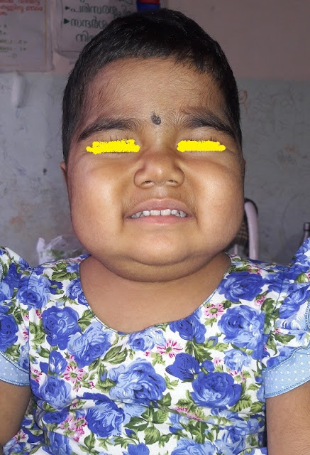
Skin and mucosal bleeds present. Caries teeth with bleeding gums.
No skeletal malformations or abnormal appearance ( This point is important in case. In view of non response always better to have a rethinking, inherited thrombocytopenias may be missed. Absent thumb, lobster thumb, triphalngeal thumb, absent radius. Spine and foot anomalies if present argue for these possibilities )
http://www.indianpediatrics.net/dec2005/dec-1246-1247.HTM
Clinical features supporting SLE and other vasculitis were not present ( HIV and SLE are to be considered in all cases like this. Girl child, especially if adolescent consider underlying SLE even when diagnostic criteria is not met )
Abdomen examination- No liver or spleen enlargement. No other palpable mass.
Other system examination were normal

Bone marrow was revised and was suggestive of ITP. She was put on full course of steroid again. She responded for a short period but became symptomatic again.
She was referred to CMC for work up and they did bone marrow as suggestive of ITP .
ANA, HIV, HbsAg negative .
She was put on Dapsone 1mg /kg which she continued for one year.
Since then she was admitted many times with symptomatic bleeds. Throughout platelets remained around 20000/mm3. Hemoglobin and RBC parameters were depending on the severity of bleed. WBC count and morphology remained normal.
Other managements tried in sequence.
All the other measures for mucosal bleeds like tranexamic acid, other supportive measures were being given during episodes.
Now she is six and half years more than two years since the onset
In January 2016, she was admitted with intracranial bleeds in the ICU . Multiple platelet transfusion done did not raise the platelet count . here one point could nt be answered. He was not given platelet transfusion for these episodes. Usually patients transfused frequenly with platelets develop antibodies so that we wont get anticipated rise of platelets with platelet transfusion .
Only option left was to do a splenectomy. But surgical risk was very high .But we had no other choice.
Paediatric surgeons did surgery with full support from paediatric team, platelet concentrate infused immediately before surgery and during surgery. Almost 10 platelet rich plasma transfused.
Surgery completed without problem, her count raised and the bleeding manifestations controlled and there was rise in platelet count following surgery.

She was discharged . Azathioprine, dapsone were continued .
Tab mycophenolate mofetil was started daily 250mg for two months and later tacrolimus (dose and duration ?) both with low dose steroids. As the condition remained same, she was referred to us.
There is no past history of medications, or significant medical illness . She is immunized up to date. No family history of excessive bleeding tendency in family for three generations.
Analysis of such a case. Few learning points
Common complaints of a case with excessive bleeding are superficial bleed in skin or mucosal bleed and occasionally deeper bleeds like GI, hematuria, IC bleed or hematoma or hemarthrosis. In the step wise clinical evaluation of these cases, we need to asses the bleeding with the following questions :1.Is it a bleed at all?
Why this question ?Occasionally drug rash like fixed drug eruption, a pigmented scar may look like a fading ecchymotic patch.
If the answer is 'yes', it is bleed.
Is this bleeding explainable by a local cause?
Just because bleeding occurs repeatedly, many cases are investigated as bleeding tendency. If it is local cause, you have to tackle the local problem responsible for the bleeding rather than doing work up of bleeding tendency.
For example, recurrent epistaxis form same side of nose may be due to epistaxis digitorum(Little's area bleeding). Patient presenting with hematemesis consider upper GI pathology as reasons eg: peptic ulcer, portal hypertension. Rule out them by clinical or appropriate investigations before asking for coagulation study or platelet workup.
If bleeding is from two or more anatomically unrelated sites, we are dealing with a case with excessive bleeding tendency. Some common mistakes which can happen when we analyse a case is epistaxis, spitting blood, hemetemasis and later malena. Just because bleeding is from multiple sites, we may decide on investigating. Here, anatomical continuity bleeding at one site may mislead you. A problem in the nose may lead on to all the above and our detailed work up will be a waste . Occasionally child abuse patient may come with skin bleeds, hematuria, hemarthrosis and mislead you .
If you ask back, "If the bleeding is from one site only, can I decide the bleeding is unlikely to be having a bleeding tendency ?" - Not always .
Bleeding on one site, we usually consider as a local problem. But, if the bleeding is out of proportion to the injury we should consider underlying bleeding tendency even if it is occurring from single site.
So, what about this case? She had bleeding from skin in the form of ecchymosis, petechiae, purpura, mucosal bleed from lips, gums and epistaxis. It cannot be explained by a local problem, she is having a bleeding tendency .
What is the pattern of bleeding ?
Is it suggestive of primary or seconday hemostasis ?
All of us know how bleeding stops following injury. First step is vasospasm and platelet plug formation. Vascular factors, platelets and Von Willebrand factor are needed for this stage (primary hemostasis ). Once this stage is over, true coagulation process sets in (secondary hemostasis). Pattern of bleeding in primary and secondary hemostasis is different which helps us in clinical evaluation to categorize these two major groups.
A bleeding in to the joint, deep hematoma occur only in coagulation disorder. It wont occur in primary hemostatic disorder.
Bleeding pattern of primary hemostatic disorder is skin, mucosal bleed, petechie, pupura and echymosis. Hematuria, GI bleeds, inracranial bleed etc can occur in severe cases of primary and secondary hemostatic disorder.
One useful point about skin bleed is to look for petechiae. Pin point bleeds will not occur in coagulation disorders. Ecchymosis can occur in both. So most useful points are bleed into a joint which definitely suggest coagulation problem and petechie which argue for primary hemostatic problem .
Occasionally we get a combination pattern of joint bleed and petechie eg in Von Willebrands's disease.
In our case, she did not have joint bleeds. She has mainly skin and mucosal bleeds, she has petechie . So we are dealing with a case of primary hemostatic problem ie vascular, platelet number, platelet function or remote possibility of Von Willebrands .
So we are half way through the analysis. It is most likely a primary hemostatic problem
Is this problem inherited one or an acquired one ?
Late onset of problem argues for an acquired condition, But that is not always the case. Occasionally milder degrees of problem may not manifest. They may manifest during a severe injury or or abnormal investigation result before preparation for surgery.One more helpful point to answer this question is positive family history. Family history is helpful in two ways.
1. It argues for a hereditary disorder
2. Constructing pedigree chart helps to indicate the pattern of inheritance and hence the type of disorder .
Classical example

So at the end of these four steps, we can narrow down in to six major group of possibilities.
Disorder of primary hemostasis - Inherited or Acquired.
Disorder of Secondary hemostasis - Inherited or Acquired.
Combination - Inherited or Acquired .
Examples of inherited primary hemostasis - hereditary thrombocytopenias, platelet function disorders, Von Willebrand's disease
Examples of acquired primary hemostasis - ITP, hematologic malignancies, SLE, disorders with small vessel vasculitis, drugs interfering with platelet function
Examples of inherited secondary/coagulation disorder - various inherited factor deficiencies
Examples of acquired coagulation disorders. liver disorders, drugs
Examples of inherited disorders of combination -Von Willebrand's
Examples of acquired disorders with combination - DIC
Most likely, our case belongs to the acquired primary hemostatic disorder. Inherited disorders of primary hemostatic disorders like inherited thrombocytopenias and platelet function disorders may present this way. But, late onset and severe manifestations from that point of time onwards. If the late manifestation is explained by mild nature it should behave as mild disorder always. In this case it is not so. So, inherited disorders less likely.
Once we have narrowed down to one of these group, try to further analyse to pin point the problem .
What are the possibilities in this group ?
1. Acquired thrombocytopenias2. Aquired vascular lesions
3. Acquired platelet function disorder
(in adults rarely acquired problems with Von Willebrand's factor occur in certain malignancies )
- We can rule out the last two easily. Acquired platelet function disorders usually occur due to drugs , toxins systems,dysfunctions like uremia etc
- Nothing supporting these possibilities are present in this case. Vascular disorders of acquired nature, vasculitis ?
- Here it is not palpable purpura. no other features of a vasculitic disorder.
- So what remains is a number problem, decrease number of platelets.
1. Is it due to decreased production from marrow ?
2. Is it due to increased destruction ?
Decreased number from bone marrow, we know of malignancies leukemia , lymphoma and rarely other ones invading bone marrow like neuroblastoma OR bone marrow aplasia .
Is it one of them? - "No "
Why ?
Total duration is more than for 2 years till now. Without definitive treatment which malignancy will survive that duration ?OK agreed. That question is easily answered here because the duration is long. Suppose you see the case at the onset of the problem? This point of duration will not help you there.
But, one point is helpful here. Look at the other blood elements- RBC and WBC. There is no evidence of involvement of RBC or WBC. Most of the time, her hemoglobin was well maintained and peripheral smear repeatedly reported their morphology as normal .
OK . If so why can't this be a selective platelet formation problem ?
Decreased production of platelet alone from bone marrow ?
Yes, it is possible. But it is rare.
So what remains is increased destruction of platelets .
What are the reasons for increased destruction? One major possibility which comes to our mind is ITP. Other rarer reasons like mechanical destruction eg microangiopathy, heart valves and other immune mediated destruction like SLE, EVANS syndrome .
Commonest among these is ITP. Thought of the above possibilities, but features supporting them were not there.
ITP .most likely. But why she has persistent bleeding and thrombocytopenia in spite of all the treatment even after two years.
How common is chronic ITP in this young kids? Chronicity is more and more common as age advances)
Coming back to our Case:
O/EVitals stable, No pallor, No jaundice , No significant lymph nodes.
(A patient with bleeding tendency may get admitted in our ICU with unstable state .Circulatory status and sensorium are the most important vitals in such cases, severe bleed or an internal bleed can cause shock.
A bleed not significant enough to cause volume loss but causing problems due to the location eg bleed in to the CNS or eyes can cause major problems. Now answer me, if his BP is high and pulse shows bradycardia, what possibility do you consider? Which physical sign will you check?).
*Features of raised intra cranial tension, level of sensorium and pupils.
(All these situations are emergencies, to be addressed without delay.)
Skin and mucosal bleeds present. Caries teeth with bleeding gums.
No skeletal malformations or abnormal appearance ( This point is important in case. In view of non response always better to have a rethinking, inherited thrombocytopenias may be missed. Absent thumb, lobster thumb, triphalngeal thumb, absent radius. Spine and foot anomalies if present argue for these possibilities )
http://www.indianpediatrics.net/dec2005/dec-1246-1247.HTM
Clinical features supporting SLE and other vasculitis were not present ( HIV and SLE are to be considered in all cases like this. Girl child, especially if adolescent consider underlying SLE even when diagnostic criteria is not met )
Abdomen examination- No liver or spleen enlargement. No other palpable mass.
Other system examination were normal

Bone marrow was revised and was suggestive of ITP. She was put on full course of steroid again. She responded for a short period but became symptomatic again.
She was referred to CMC for work up and they did bone marrow as suggestive of ITP .
ANA, HIV, HbsAg negative .
She was put on Dapsone 1mg /kg which she continued for one year.
Since then she was admitted many times with symptomatic bleeds. Throughout platelets remained around 20000/mm3. Hemoglobin and RBC parameters were depending on the severity of bleed. WBC count and morphology remained normal.
Other managements tried in sequence.
- IV gamma globulin,
- Azathioprine 1.5 mg /kg,
- Anti D 75 mg/kg
- and Pulse Dexona 20 mg/m2 monthly.
All the other measures for mucosal bleeds like tranexamic acid, other supportive measures were being given during episodes.
Now she is six and half years more than two years since the onset
In January 2016, she was admitted with intracranial bleeds in the ICU . Multiple platelet transfusion done did not raise the platelet count . here one point could nt be answered. He was not given platelet transfusion for these episodes. Usually patients transfused frequenly with platelets develop antibodies so that we wont get anticipated rise of platelets with platelet transfusion .
Only option left was to do a splenectomy. But surgical risk was very high .But we had no other choice.
Paediatric surgeons did surgery with full support from paediatric team, platelet concentrate infused immediately before surgery and during surgery. Almost 10 platelet rich plasma transfused.
Surgery completed without problem, her count raised and the bleeding manifestations controlled and there was rise in platelet count following surgery.

She was discharged . Azathioprine, dapsone were continued .
But that response did not last long. She started to have skin and mucosal bleeds again. Platelet counts dropped again.
Why did this happen? She showed initial response but gradual worsening again, is it accessory spleen?
Peripheral smear was checked for the evidence of splenectomy. Peripheral smear did not show changes which we expect in splenectomy. So possibility of accessory spleen was high .
Why did this happen? She showed initial response but gradual worsening again, is it accessory spleen?
Peripheral smear was checked for the evidence of splenectomy. Peripheral smear did not show changes which we expect in splenectomy. So possibility of accessory spleen was high .
An ultra sonogram abdomen to look for accessory spleen did not show any. Accessory spleen may not be picked up in USG. May be CT or nucleotide scan will give better results.But was not done.
She showed better response following pulse Dexamethazone.
She is now admitted for pulse Dexamethazone , Azathioprine dose increased to 2 mg/kg .
What next ?
She is now admitted for pulse Dexamethazone , Azathioprine dose increased to 2 mg/kg .
What next ?
We are planning a trial with with Rituximab. Of course cost is an issue.
Follow up
She was given a course of Rituximab . Doing well after six months now . Azathiprim and Dapsone is continued .
Now she is admitted with pneumonia

Follow up
She was given a course of Rituximab . Doing well after six months now . Azathiprim and Dapsone is continued .
Now she is admitted with pneumonia


No comments:
Post a Comment